Campbell, Amy, Franco, Catarina Ferraz, Su, Ling-I, Perkins, Simon, Jones, Andrew  ORCID: 0000-0001-6118-9327, Brownridge, Philip, Perkins, Neil and Eyers, Claire ORCID: 0000-0002-3223-5926
(2020)
Temporal modulation of the NF-κB RelA network in response to different types of DNA damage.
[Preprint]
ORCID: 0000-0001-6118-9327, Brownridge, Philip, Perkins, Neil and Eyers, Claire ORCID: 0000-0002-3223-5926
(2020)
Temporal modulation of the NF-κB RelA network in response to different types of DNA damage.
[Preprint]
![[img]](https://livrepository.liverpool.ac.uk/style/images/fileicons/text.png) |
Text
Campbell et al. RelA resubmission 11Jan2020_supplementary material .docx - Author Accepted Manuscript Download (30kB) |
|
Text
34966_1_merged_1610378043.pdf - Author Accepted Manuscript Download (1MB) | Preview |
|
|
Text
Supplementary figure 1_11Jan2021.pdf - Author Accepted Manuscript Download (390kB) | Preview |
|
![[img]](http://livrepository.liverpool.ac.uk/3113415/17/Supp%20Fig%202%20A%20gif%20significant%20changes.gif) |
Image
Supp Fig 2 A gif significant changes.gif - Author Accepted Manuscript Download (2MB) | Preview |
![[img]](http://livrepository.liverpool.ac.uk/3113415/22/Supp%20Fig%202%20B%20Time%20dependent%20CE.gif) |
Image
Supp Fig 2 B Time dependent CE.gif - Author Accepted Manuscript Download (100MB) | Preview |
|
Image
Supp Fig 3.tif - Author Accepted Manuscript Download (327kB) | Preview |
|
![[img]](https://livrepository.liverpool.ac.uk/style/images/fileicons/other.png) |
Spreadsheet
Table S1_Supplementary_Network_KmeansClusters.xlsx - Author Accepted Manuscript Download (454kB) |
|
Spreadsheet
Tables S2_S3 ETO_HU FinalTable_CE v2.xlsx - Author Accepted Manuscript Download (3MB) |
Abstract
<h4>ABSTRACT</h4> Different types of DNA damage can initiate phosphorylation-mediated signalling cascades that result in stimulus specific pro- or anti-apoptotic cellular responses. Amongst its many roles, the NF-κB transcription factor RelA is central to these DNA damage response pathways. However, we still lack understanding of the co-ordinated signalling mechanisms that permit different DNA damaging agents to induce distinct cellular outcomes through RelA. Here, we use label-free quantitative phosphoproteomics to examine the temporal effects of exposure of U2OS cells to either etoposide (ETO) or hydroxyurea (HU) by monitoring the phosphorylation status of RelA and its protein binding partners. Although few stimulus-specific differences were identified in the constituents of phosphorylated RelA interactome after exposure to these DNA damaging agents, we observed subtle, but significant, changes in their phosphorylation states, as a function of both type and duration of treatment. The DNA double strand break (DSB)-inducing ETO invoked more rapid, sustained responses than HU, with regulated targets primarily involved in transcription, cell division and canonical DSB repair. Kinase substrate prediction of ETO-regulated phosphosites suggest abrogation of CDK1 and ERK1 signalling, in addition to the known induction of ATM/ATR. In contrast, HU-induced replicative stress mediated temporally dynamic regulation, with phosphorylated RelA binding partners having roles in rRNA/mRNA processing and translational initiation, many of which contained a 14-3-3ε binding motif, and were putative substrates of the dual specificity kinase CLK1. Our data thus point to differential regulation of key cellular processes and the involvement of distinct signalling pathways in modulating DNA damage-specific functions of RelA.
| Item Type: | Preprint |
|---|---|
| Uncontrolled Keywords: | dna damage, etoposide, hydroxyurea, mass spectrometry, phosphoproteomics, phosphorylation, Rel A |
| Depositing User: | Symplectic Admin |
| Date Deposited: | 13 Jan 2021 09:19 |
| Last Modified: | 18 Jan 2023 23:03 |
| DOI: | 10.1101/2020.08.11.246504 |
| Related URLs: | |
| URI: | https://livrepository.liverpool.ac.uk/id/eprint/3113415 |
 Dimensions
 Dimensions
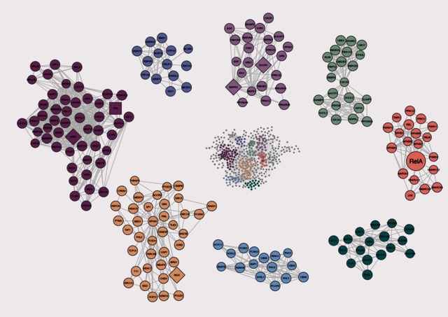

